Abstract:
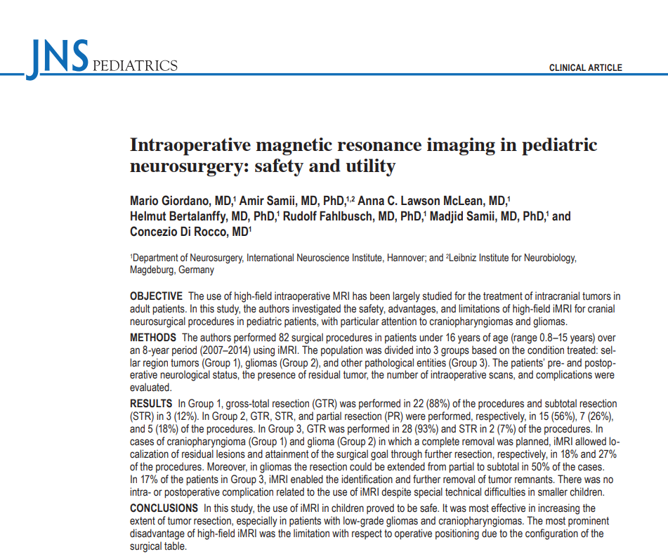
The use of high-field intraoperative MRI has been largely studied for the treatment of intracranial tumors in adult patients.In this study,the authors investigated the safety,advantages,and limitations of high-field iMRI for cranial neurosurgical procedures in pediatric patients,with particular attention to craniopharyngiomas and gliomas.METHODS The authors performed 82 surgical procedures in patients under 16 years of age(range 0.8-15 years)over an 8-year period using iMRI.The population was divided into 3 groups based on the condition treated:sellar region tumors(Group 1),gliomas(Group 2),and other pathological entities(Group 3).The patients'pre-and postoperative neurological status,the presence of residual tumor,the number of intraoperative scans,and complications were evaluated.RESULTS In Group 1,gross-total resection(GTR)was performed in 22(88%)of the procedures and subtotal resection(STR)in 3(12%).In Group 2,GTR,STR,and partial resection(PR)were performed,respectively,in 15(56%),7(26%),and 5(18%)of the procedures.In Group 3,GTR was performed in 28(93%)and STR in 2(7%)of the procedures.In cases of craniopharyngioma(Group 1)and glioma(Group 2)in which a complete removal was planned,iMRI allowed localization of residual lesions and attainment of the surgical goal through further resection,respectively,in 18%and 27%of the procedures.Moreover,in gliomas the resection could be extended from partial to subtotal in 50%of the cases.In 17%of the patients in Group 3,iMRI enabled the identification and further removal of tumor remnants.There was no intra-or postoperative complication related to the use of iMRI despite special technical difficulties in smaller children.In this study,the use of iMRI in children proved to be safe.It was most effective in increasing the extent of tumor resection,especially in patients with low-grade gliomas and craniopharyngiomas.The most prominent disadvantage of high-field iMRI was the limitation with respect to operative positioning due to the configuration of the surgical table.

中文摘要:术中高场强磁共振成像在成人颅内肿瘤的治疗中已经得到了广泛的研究。在这项研究中,作者调查了高场强磁共振成像在儿童颅神经外科手术中的顺利性、优势和局限性,特别是颅咽管瘤和胶质瘤。方法作者对82例16岁以下(0.8-15岁)的患者进行了82次手术。根据治疗情况将人群分为3组:鞍区肿瘤(1组)、胶质瘤(2组)和其他病理实体(3组)。评估患者术前和术后的神经系统状况、肿瘤残留情况、术中扫描次数和并发症。结果1组22例(88%)行全切除,3例(12%)行次全切除。2组分别有15例(56%)、7例(26%)和5例(18%)行GTR、STR和部分切除(PR)。3组28例(93%)行GTR,2例(7%)行STR。对于计划完全切除的颅咽管瘤(1组)和胶质瘤(2组),在18%和27%的手术中,iMRI允许定位残余病灶并通过进一步切除达到手术目的。此外,在胶质瘤中,50%的病例可以从部分切除扩大到次全切除。在3组中,17%的患者能够识别并进一步清除肿瘤残余物。尽管在较小的儿童中有不同的技术困难,但没有出现与使用iMRI相关的术中或术后并发症。所以儿童应用iMRI是顺利的。对提高肿瘤切除范围合适,是对低度胶质瘤和颅咽管瘤患者。高场iMRI突出的缺点是由于手术台的结构限制了手术定位。
简介:术中磁共振成像(iMRI)的引入是颅内肿瘤外科治疗的一项重要创新。低场磁共振成像的一开始报告可以追溯到20世纪90年代中期。在随后的几年里,手术方案和磁共振成像技术不断发展,使得高场扫描仪得以用于术中成像。场强越大,信噪比越高,成像质量越好。这一发展导致了一个更广泛的潜在应用,即通过描绘肿瘤与周围功能相关结构的空间关系来提高肿瘤检测的准确性和提高肿瘤切除的顺利性。目前,磁共振成像(iMRI)是一种成熟的工具,它结合了基于术前和术中图像的计算机神经导航技术,可以顺利、完整地切除成人脑胶质瘤。除了一些早期的初步报告,iMRI在儿科人群中的应用仅在较近的几项研究中进行过调查。对于儿童来说,肿瘤切除的程度至关重要,因为它是颅内恶性肿瘤的主要预后因素,颅内良性肿瘤完全切除是可以完全治愈的,而术后出现意外的肿瘤残留可能导致再次手术。在这篇报告中,我们介绍了我们在儿科人群中应用高场强磁共振成像的经验。我们将讨论这项技术的顺利性和实用性,并介绍患者的短期预后。
方法:作者在8年的时间里,使用iMRI对16岁以下(0.8-15岁)的患者进行了82次外科手术。根据治疗情况将人群分为3组:鞍区肿瘤(1组)、胶质瘤(2组)和其他病理实体(3组)。评估患者术前和术后的神经系统状况、肿瘤残留情况、术中扫描次数和并发症。

结果:1组22例(88%)行全切除术,3例(12%)行次全切除术。2组分别有15例(56%)、7例(26%)和5例(18%)行GTR、STR和部分切除(PR)。3组28例(93%)行GTR,2例(7%)行STR。对于计划完全切除的颅咽管瘤(1组)和胶质瘤(2组),在18%和27%的手术中,iMRI允许定位残余病灶并通过进一步切除达到手术目的。此外,在胶质瘤中,50%的病例可以从部分切除扩大到次全切除。在3组中,17%的患者能够识别并进一步清除肿瘤残余物。
结论:在这项研究中,iMRI在儿童中的应用被证明是顺利的。对提高肿瘤切除范围合适,是对低级别胶质瘤和颅咽管瘤患者。高场iMRI比较突出的缺点是由于手术台的结构限制了手术定位。
原文链接:https://www.sci-hub.pl/10.3171/2016.8.peds15708


